Advances in mammography and breast imaging

Home
Views
Information
Research
Links
Site map

This site is an EPSRC funded public awareness project
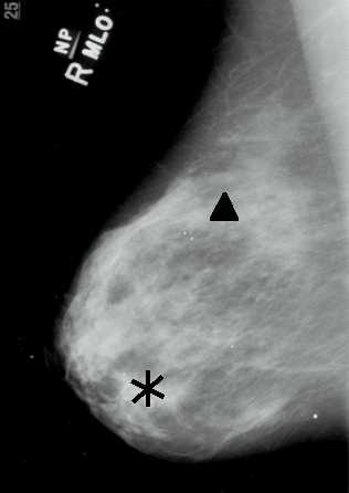
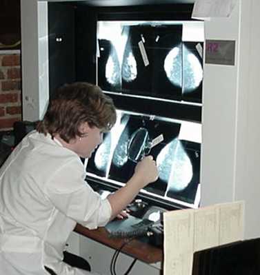
Computer-aided detection

What is CAD?
Benefits of CAD
Limitations of CAD
History of CAD


What is CAD?

Computer-aided detection (CAD) is a recent advance in mammography which helps to identify abnormalities within the breast. CAD technology works by reviewing digitised mammograms and marking areas of suspected abnormalities. The radiologist then reviews whether the marked areas are suspicious and require additional imaging tests or biopsy. When using a CAD system the radiologist always makes the final interpretation of the mammogram.

Mammogram with prompts Computer software detects suspected abnormalities and marks microcalcifications with a triangle and masses with an asterisk.

ImageChecker A radiologist reviews the marked areas, referring back to the original mammogram.

Benefits of CAD

  • It can enhance the radiologist's performance by drawing attention to suspicious areas that may have been missed by the radiologist.
  • It can improve the rate of detection of small breast abnormalities which increases the chances of successful treatment.
  • It performs consistently, reducing variability due to fatigue, distraction and workload.
  • It reduces site-to-site variability.

Limitations of CAD

  • It currently requires a lot of time and effort to digitise mammogram films, however the increased use of digital mammography will, in time, increase its efficiency.
  • It currently marks a significant number of normal areas on mammograms as abnormal, which could lead to unnecessary additional breast imaging and biopsies and distracts the reporting radiologist.
  • There is currently only one CAD device commercially available and it is very expensive.

History of CAD

1967 Dr Fred Winsberg and co-workers were amongst the first published investigators to considered the difficulty of viewing large number of screening mammograms.
1970s Researchers begin using computers to extract properties of lesions including calcification, spiculation, roughness and shape. These properties are later termed features and the number of features has since substantially expanded.
1990s Computers have become more powerful and can process digital images more quickly.
1998 The first commercially available CAD system is introduced.

Mammography | Digital mammography | CAD | MRI | Ultrasound | Glossary